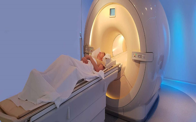
In medicine, exciting times are now. Industry disruptors like gene editing, 3D printing, and artificial intelligence have made their way into the mainstream of medical care during the past ten years. Additionally, diagnostic technology is developing quickly. X-rays, MRIs, CT scans, PET scans, mammograms, and ultrasounds are just a few of the medical imaging studies that are currently used to diagnose or confirm disease, injury, or abnormalities. However, each of these scans’ degree of clarity and resolution is not always ideal, and new, creative imaging technologies intended to enhance patient diagnoses and outcomes are always being developed.
1. Artificial Intelligence And Machine Learning (AI And ML)
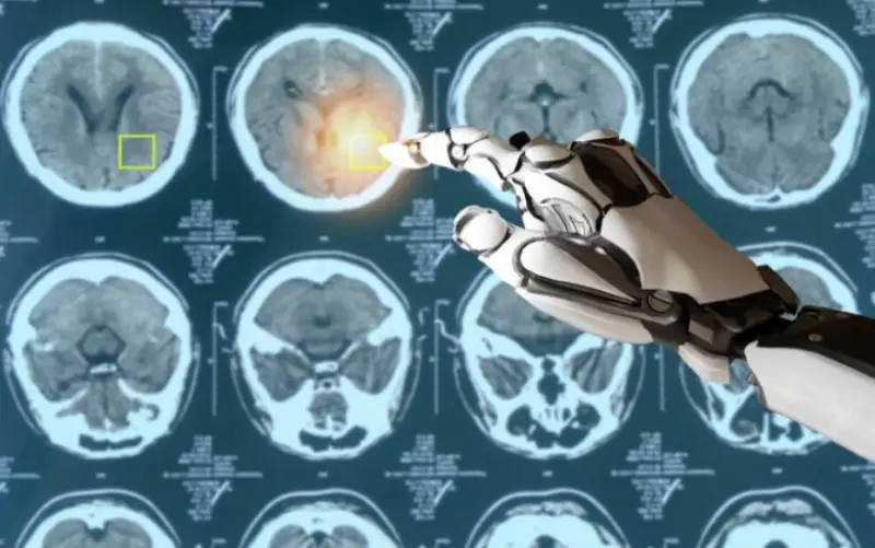
The radiological workflow has recently been improved by AI and ML utilizing more sophisticated detection techniques. A massive stroke can be immediately identified by AI, which can then direct the scan to the top of the radiologist’s imaging stack for faster diagnosis and emergency treatment within a constrained therapeutic window. For radiologists who review large numbers of scans each day, AI and ML also increase procedural efficiency on a day-to-day basis. Deep-learning algorithms may soon be used by AI and ML to mine data for novel illness indicators.
2. Photon Counting Detector (PCD) Technology
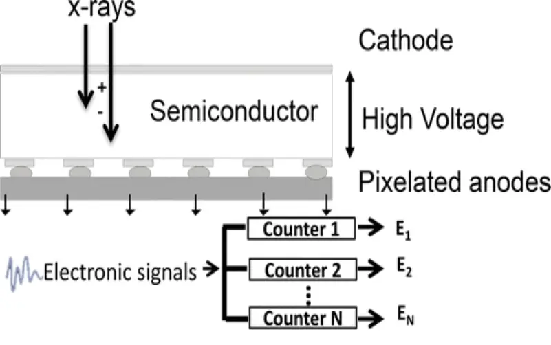
After each X-ray has gone through a scan area, PCD, a kind of computed tomography (CT) technology, sorts the energy from each one. With PCD, numerous sets of CT data may be produced from a single X-ray with greater visualization accuracy and finer segmentation than what radiologists are accustomed to seeing in typical CT scan pictures. In point-of-care emergency and surgical circumstances, where fast access to high-accuracy imaging might save a life, a mobile PCD CT would be very beneficial.
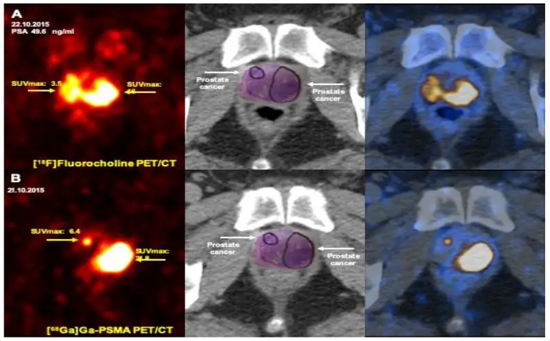
3. Prostate-specific Membrane Antigen (PSMA) PET Scans
PSMA PET scans are expected to replace traditional methods of identifying prostate cancer and spotting its metastases as the new gold standard of treatment by 2022, according to the Cleveland Clinic’s list of the top 10 medical innovations to watch. On cancer surface cells, the prostate cancer biomarker PMSA is present in significant concentrations. The most prevalent type of cancer in males, particularly among older men, is prostate cancer, which affects 200,000 people in the United States each year. Current MRI and CT scans for prostate imaging have limitations in terms of precision and clarity. Improved treatment and results for men with prostate cancer may result from this cutting-edge technique that involves the use of radioactive tracers on PMSA proteins and the visualization of the cells using CT and MRI.
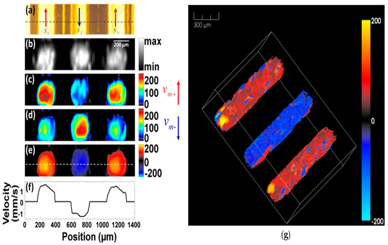
4. Electromagnetic Acoustic (EMA) Imaging
Currently, ultrasounds alone (ultrasounds only employ sound waves) are unable to create precise, real-time visualizations of fluid buildup, calcification, diseased tissue, and capillaries. EMA mixes radio signals with sound waves to do this. EMA differs from existing medical imaging scans essentially: it may be utilized immediately at the point of care (during a doctor’s appointment) to correctly picture and diagnose a medical issue. This avoids the need for a second referral to an imaging facility and the stressful procedure of anticipating your findings.
5. 3D Mammography
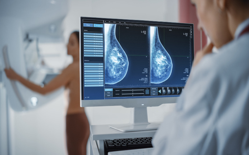
Since many years ago, digital breast tomosynthesis (DBT) systems have been gradually replacing conventional 2D mammography units, and by 2022, their share of breast imaging centers will be close to 50%. Traditional 2D preventative mammograms use X-rays to make pictures from two different angles, but 3D mammography devices operate more like computed tomography (CT) scans, taking a large number of slice-by-slice images that are combined to produce a composite image. Although they are pricey, they make it easier for radiologists to distinguish between dense breast tissue and cancerous tissue. Additionally, they tend to lessen the number of biopsies and follow-up appointments needed by women following their diagnostic or preventative screenings.
6. 3D Lung X-rays
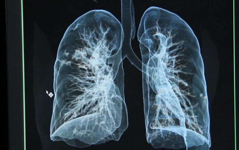
A high-definition, 3D X-ray picture of lungs breathing in real-time is now being tested in Sweden. The smallest and most minute structures in the lungs of mice have already been observed by scientists during inhalation and exhalation by the mice. This is significant since the patient must stay motionless for the majority of imaging studies to get quality pictures. Here, breathing naturally does not degrade the sharpness of the picture. Once authorized, this imaging technology poses little risk to people because it only uses a very modest dosage of radiation.
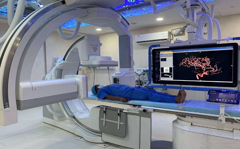
7. Interventional Radiology
Interventional Radiology is a medical subspecialty that uses a minimally invasive, image-guided approach to diagnose and treat almost every organ system. Interventional radiology’s goals are to reduce patient risk and enhance health outcomes. In-depth understanding of minimally invasive treatments employing needles, wires, and catheters to administer therapies through a pinhole with low morbidity and quicker recovery times than traditional surgical procedures is provided by the interventional radiology technique.
8. Functional Magnetic Resonance Imaging
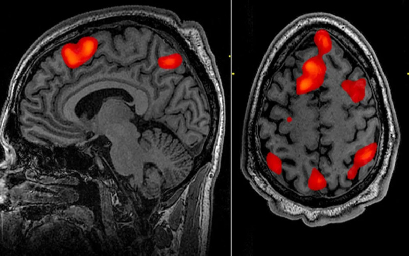
The fMRI technique tracks minute variations in blood flow that are brought on by brain activity. It may be used to determine which brain regions are in charge of crucial functions, assess the consequences of a stroke or another illness, or direct brain treatment. Other imaging methods may be unable to find abnormalities in the brain that fMRI can.
9. Optical Coherence Tomography (OCT)
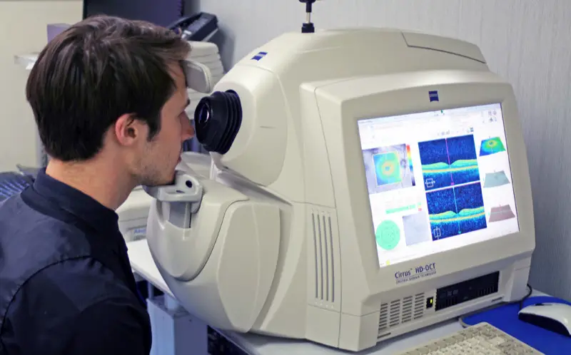
Non-invasive imaging procedures include optical coherence tomography (OCT) and optical coherence tomography angiography (OCTA). They photograph your retina in cross-section using light waves. Your ophthalmologist can view each of the different layers of the retina using OCT. Your ophthalmologist can map and gauge their thickness in this way. These actions support the diagnosis. They also oversee the treatment of glaucoma, age-related macular degeneration (AMD), diabetic eye disease, and other retinal disorders.
10. Computed Tomography (CT)
The term “computed tomography,” or CT, refers to a computerised X-ray imaging technique in which a patient is exposed to a focused X-ray beam that is rapidly rotated around the body, creating signals that are then processed by the machine’s computer to create cross-sectional images, or “slices.” These sections, sometimes referred to as tomographic photos, can give a doctor more detailed information than standard X-rays. Once the computer of the machine has collected multiple sequential slices, a three-dimensional (3D) image of the patient may be produced. The underlying anatomy of the patient as well as any prospective tumours or abnormalities are easier to spot as a result.
